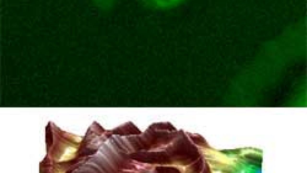
UC Davis researchers in nanotechnology, chemistry and biology now have access to one of the most advanced microscopes of its type in the world. The new Spectral Imaging Facility, opened this fall, is a combination of an atomic force microscope and a laser scanning confocal microscope, the first commercial machine of its kind.
The atomic force microscope was built by Asylum Research of Santa Barbara and the confocal microscope system by Olympus America Inc. Integration of the two systems was carried out by Asylum Research with the participation of scientists led by Gang-yu Liu, professor of chemistry at UC Davis. Acquisition of the instrument was funded by a grant of $354,000 from the National Science Foundation, matched by $151,714 from UC Davis.
The confocal microscope allows three-dimensional imaging of samples, such as cells, and picks up structures tagged with fluorescent dyes. Instead of cutting a sample into thin slices, a researcher can focus through the entire sample and resolve it in three dimensions.
The atomic force microscope uses an extremely fine tip to run over the surface of a sample and "see" extremely fine detail down to an atomic scale.
"It allows you to see both the detail and the bulk," Liu said.
For example, biologists could use fluorescent tags to look at structures inside a cell and link them to very small changes on the cell membrane. Materials scientists could use it to get information about the bulk structure of a material and to measure the arrangement of atoms at the surface. The tip of the atomic force microscope can also be used as a probe to nudge cells, or place atoms or molecules into new, microscopic patterns.
The project includes 24 UC Davis faculty from the departments of Chemistry and of Physics; the colleges of Engineering, Agricultural and Environmental Sciences, and Biological Sciences; the School of Medicine; and from the Lawrence Livermore National Laboratory.
Media Resources
Andy Fell, Research news (emphasis: biological and physical sciences, and engineering), 530-752-4533, ahfell@ucdavis.edu
Gang-yu Liu, UC Davis Department of Chemistry, (530) 754-9678, liu@chem.ucdavis.edu
